Identifying the Cricothyroid Membrane with String of Pearls

Cricothyroid Membrane Identification in Sagittal Scan
In the sagittal plane, the thyroid cartilage will move from superficial to deep as it moves inferiorly until its border with the CTM. The cricoid cartilage will appear inferior to the CTM as a hypoechoic ovoid structure, and the tracheal rings will appear as smaller evenly spaced intermittent "pearls" or "beads on a string". Tc, thyroid cartilage; Cc, cricoid cartilage. Very high utility scan especially in marking the tracheostomy incision site.
Longitudinal Approach ("String of Pearls"): The probe is placed lengthwise in the midline; tracheal rings appear like a string of beads, and the distinct gap superior to the cricoid and inferior to the thyroid cartilage defines the cricothyroid membrane.
Marking the Incision Site
Using a straight pin, paper clip or IV catheter, the ultrasound is used to mark the incision site with a "sharpie" prior to incision by creating dropout from the object on the field. Center the CTM precisely on the US screen, then mark the exact puncture site.
Verification of Intubation
Visualization of the ET tube
The endotracheal tube is visualized with either a "double tract" characteristic or filling the cuff with saline. First the trachea or cricoid cartilage is visualized using transverse and sagittal plane. The esophagus should also be interrogated for the ET tube along with "lung sliding" which is evidence of lung inflation and deflation.
Lung Sliding
Lung sliding ("ants marching on a string") is a sign of correct ET tube placement. However, if the tube is too deep (mainstem intubation) there may be selective sliding. As well pleurodesis may affect sliding such that this may need to be assessed for.
Redundant Exam?
In the "Tool or Toy" category, I am gong to go out on a limb and say this may be a redundant scan. I am sure the senior clinician who took the image of the ET tube in the esophagus knew where it was. Regardless the empty esophagus appears as a flat, multi-layered structure usually situated to the left of the trachea. Seeing a tube inside this structure confirms an esophageal intubation The "Double Track" or "Sign of the Ring": When a tube enters the esophagus instead of the trachea, the scan shows a dilated, rounded structure next to the trachea containing a distinct double hyperechoic line or air artifact (often called the double-track sign)
Gastric Ultrasound Technique
Intro to Gastric Ultrasound
Gastric ultrasound is used when prandial status is uncertain or gastric emptying may be delayed. Images are acquired in the epigastrium in the right lateral decubitus position. The liver is the best point of reference, and visualization of the aorta and superior mesenteric artery help define the correct plane. The gastric antrum may be empty or contain fluids or solid food. If fluid, quantification of the volume will dictate the risk and airway management.
Interpretation
Once the images are obtained, decision-making involves content and volume. When the stomach is empty, it appears small, flat, and collapsed in both the supine and RLD positions and has been described as having a ‘bulls-eye’ appearance. Gastric secretions and clear fluids, such as water, clear juices, or black tea, are anechoic or hypoechoic in appearance. Recent consumption of clear fluids or carbonated drinks can produce air bubbles within the fluid that may appear as hyperechoic dots. Solid matter are invariably associated with the ingestion of air which prevents sonographic visualization of deeper structures and creates a ‘frosted glass’ appearance with ring down artifact.l
Estimating Gastric Fluid Volume
Empty Compared to Fluid Filled Stomach
Using a BodyWorks Eve simulator, empty and fluid-filled gastric antrum scans are toggled back and forth. There s a stark difference between the bull's eye/target with the empty antrum compared to the anechoic, thin walled fluid filled antrum. If scanning with a Butterfly of other handheld/console system that allows cross sectional area no calculation is necessary. If using a system with simple calipers/distance measurements the anterior-posterior and cranial-caudal dimensions are assessed.

Measuring Fluid Volume by CSA
Using the same fluid filled scan from the simulator, the anterior-posterior and cranial-caudal dimensions are plugged into the formula for area. While not measuring ml/kg it is more simple than
Volume (ml)=27 + 14.6 (CSA)-1.28 (age) and serves the same purpose.
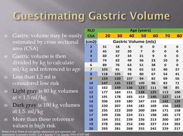
Use of CSA in Decision-making
Volume estimation based on a CSA of the gastric antrum has been the focus of multiple recent investigations. A clinically relevant ‘‘cut-off’’ value of CSA would be one that differentiates a baseline volume (Grade 0 or Grade 1 antrum or <1.5 mLkg-1) from a greater-than-baseline condition (Grade 2 antrum or >1.5 mLkg-1). This value of antral CSA could be a simple surrogate measure that could facilitate the interpretation of examination findings when clear fluid is observed in the antrum. Based on this metric a CSA less than 8 cm2 in women or 10 cm2 in men is acceptable.

Gastric Ultrasound in Decision-Making
An ‘‘empty’’ stomach (Grade 0 antrum) or a low volume of clear fluid within the range of baseline gastric secretions (Grade 1 antrum or <1.5 mLkg-1) is consistent with a fasting state and suggests a low risk for aspiration. In the absence of other risk factors, the ultrasound confirmation of an empty stomach would indicate that no special airway management precautions (intubation, rapid sequence induction) are required, and that supraglottic airway devices or deep sedation without airway protection may be appropriate management choices. Conversely, solid content or a high volume of clear fluid that is not in keeping with a fasting state suggests a higher-than-baseline risk for aspiration. Regardless this should only serve to complement the clinician's decision-making.
Lung Scans

Pulmonary/Lung Ultrasound
Point of Care ultrasound (POCUS) of the lungs, also known as lung ultrasound (LUS), has emerged as a technique that allows for pulmonary assessment with greater accuracy and speed compared to conventional techniques such as chest x-ray and auscultation. Lung ultrasound is dependent on the interpretation of ultrasound artifacts with unique signatures that allow interpretation of pathologies such as pneumothorax, pulmonary edema, effusion, pneumonia and even failed intubation. Visceral and parietal pleura appear as hyperechoic and movement or "lung sliding" represents ventilation. Consolidation and "B" lines represent pathology.
Lung Zones
Zones 1 and 2 represent the anterior lung quadrants and are most relevant for pneumothorax and interstitial edema. Zones 3 and 4 represent the lateral chest wall and are most relevant for pneumothorax, consolidation and pleural effusion. Zones 5 and 6 are posterior and represent gravity dependent pathologies such as pleural effusion and consolidation. Typically reference points for zones 4 and 6 are the liver and spleen.
Lung Pathologies
In this simulator based example we have B-lines (bright vertical laser like artifacts) that signal fluid or thickening in the lung tissue. This could represent pulmonary edema or interstitial disease as with COVID. In addition consolidation is present. Needless to say interpretation will depend on clinical presentation.